
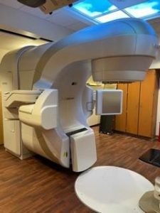
 Indiana University Health Arnett Cancer Center has added a new linear accelerator to its cancer fighting toolkit. The new linear accelerator replaces one that is roughly nine years old. With the addition of a new linear accelerator in 2019, the IU Health Arnett Cancer Center now has two state of the art Varian Truebeam machines.
A linear accelerator (LINAC) is a machine that is utilized to treat cancer by generating high energy x-rays or electrons. These x-rays or electrons can be delivered with pinpoint accuracy and are able to conform to a tumor’s shape resulting in destruction of cancer cells while sparing surrounding normal tissue.
This cutting-edge machine boasts some features and benefits not available anywhere else in the greater Lafayette area. These advances allow patients to be treated faster and with more certainty that the radiation is always targeting the cancer. A LINAC uses optimized guided radiotherapy technology to shape radiation beams to precisely match a patient’s tumor. This allows pinpoint accuracy of radiation delivery to the areas that need it, protecting surrounding tissue and reducing the risk of side effects. In addition, the sophisticated design drastically reduces a patient’s table time resulting in treatments that take as little as five minutes.
“Having the ability to deliver effective treatments in less time with a greater degree of certainty is a win for the patient and aligns perfectly with our mission to provide the best care designed for each patient,” said Matthew Orton, MD, radiation oncologist with IU Health Arnett Cancer Center. “This new technology allows us to deliver the most targeted and effective radiation treatment available anywhere.”
A LINAC is the device most commonly used for external beam radiation treatments for patients with cancer. Medical LINACs use electricity rather than a radioactive source to generate electron beams or high energy x-rays for the targeted destruction of cancer cells. It features several built-in safety measures to ensure that it will deliver the dose as prescribed and is routinely checked by a medical physicist to ensure it is working properly.
IU Health Arnett Cancer Center Advanced Treatment Capabilities
- Image-Guided Radiation Therapy (IGRT)
- Intensity-Modulated Radiation Therapy (IMRT)
- Cone Beam Computed Tomography (CBCT) – Offers real time 3D imaging for increased accuracy in treatment delivery. CBCT imaging provides enhanced views of soft tissues which can be difficult to do with standard KV/MV imaging
- Six Degrees of Freedom Treatment Couch (6DoF) – This couch is designed to adjust seamlessly on six axes, without reentering the treatment room to adjust the patient. This can greatly reduce treatment time and increase treatment accuracy.
- Stereotactic Body Radiation Therapy (SBRT) delivers extremely precise radiation treatments with greater doses per treatment and fewer overall treatments.
- Stereotactic Radiosurgery (SRS) –Delivers extremely precise radiation treatments with a large dose treatment to a small target. Generally, less than 3cm.
- Breast Brachytherapy- Interstitial breast irradiation utilizing catheter-based techniques.
- High Dose Rate (HDR) Vaginal Cuff Brachytherapy.
- Respiratory-Gated Radiotherapy-Uses a Respiratory Position Monitoring system for significant improvement in the irradiation of tumor sites affected by respiratory motion. Primarily lung, liver and breast
- Calypso Target Tracking System- utilizes electromagnetic transponders implanted inside the treatment target to ensure exact target location for patient setup and treatment. It has the ability to stop treatment automatically if a target were to move outside the treatment field during a radiation treatment.
- Optical Surface Monitoring System – This system is equipped with special cameras that create a 3D image of the patient’s body and ensures that the body contours match the exact body position for which the treatment plan was created. This allows for an extremely accurate treatment positioning and monitoring.
- CT Simulations – An onsite wide boar CT simulator allows for treatment planning CT simulations to delineate treatment target volumes and allow for sophisticated radiation treatment planning. Oral and IV contrast can be utilized to help identify target volumes.
- 4D CT Treatment Planning Simulations – combines the functions of respiratory position monitoring system with traditional CT data. This provides physicians and dosimetrist the ability to view the treatment target as it moves through a patient’s breathing pattern, ensuring that the treatment plan is created to account for tumor location during the entirety of irradiation.